Labelled Pictures Of Human Skin : The Skin Human Anatomy Picture Definition Function And Skin Conditions
Labelled Pictures Of Human Skin : The Skin Human Anatomy Picture Definition Function And Skin Conditions. Find the perfect skin cell diagram stock photos and editorial news pictures from getty images. Picture of human body with organs labeled, download this wallpaper for free in hd resolution.picture of human body with organs labeled was posted in june 11, 2017 at 5:58 am. Description from picture of human body with organs labeled pictures wallpaper : Skin is made up of two layers that cover a third fatty layer. Anatomy human body organs female 14 photos of the anatomy human body organs female anatomy human body appendix, anatomy human body liver, anatomy human body spleen, anatomy human body systems, anatomy of human body organs male, anatomy of the human body internal organs, diagram of human body organs in.
ads/bitcoin1.txt
Also explore over 33 similar quizzes in this category. Desquamation (sloughing of cells) from the epidermis, thick skin, human, 100x at 35mm. Beneath the two layers is a layer of subcutaneous fat, which also protects your body and helps you adjust to outside temperatures. This hd wallpaper picture of human body with organs labeled has viewed by 1017 users. Webmd's skin anatomy page provides a detailed image of the skin and its parts as well as a medical definition.
How To Draw The Diagram Of Human Skin Easily Youtube from i.ytimg.com Outer layer of the skin. Skin has two main layers, both of which serve a purpose. The epidermis, an outermost layer that contains the primary protective structure, the stratum corneum; Don't forget to share this picture with others via. Though nearly all human skin is covered with hair follicles, it can appear. Using the result from the previous section, we proceed to determine which regions can possibly determine a frontal human face. Minute holes from which sweat and sebum are secreted. 396 x 407 photo description:
Find free pictures, photos, diagrams, images and information related to the human body right here at science kids.
ads/bitcoin2.txt
Skin also helps maintain a constant body temperature. The cells in all of the layers except the stratum basale are called keratinocytes. The human skin is the outer covering of the body and is the largest organ of the integumentary system.the skin has up to seven layers of ectodermal tissue and guards the underlying muscles, bones, ligaments and internal organs. Beneath the two layers is a layer of subcutaneous fat, which also protects your body and helps you adjust to outside temperatures. Corneum, granulosum, spinosum, basale), dermis, sweat gland ducts and desquamating cells sloughing off the surface. Desquamation (sloughing of cells) from the epidermis, thick skin, human, 100x at 35mm. Literally covering you from head to toe. Outer layer of the skin. The outer layer is called the epidermis; Discover (and save!) your own pins on pinterest This hd wallpaper picture of human body with organs labeled has viewed by 1017 users. Skincare, bodycare, healthcare, hygiene and medicine concept. Despite being just a few millimeters thick, skin makes up.
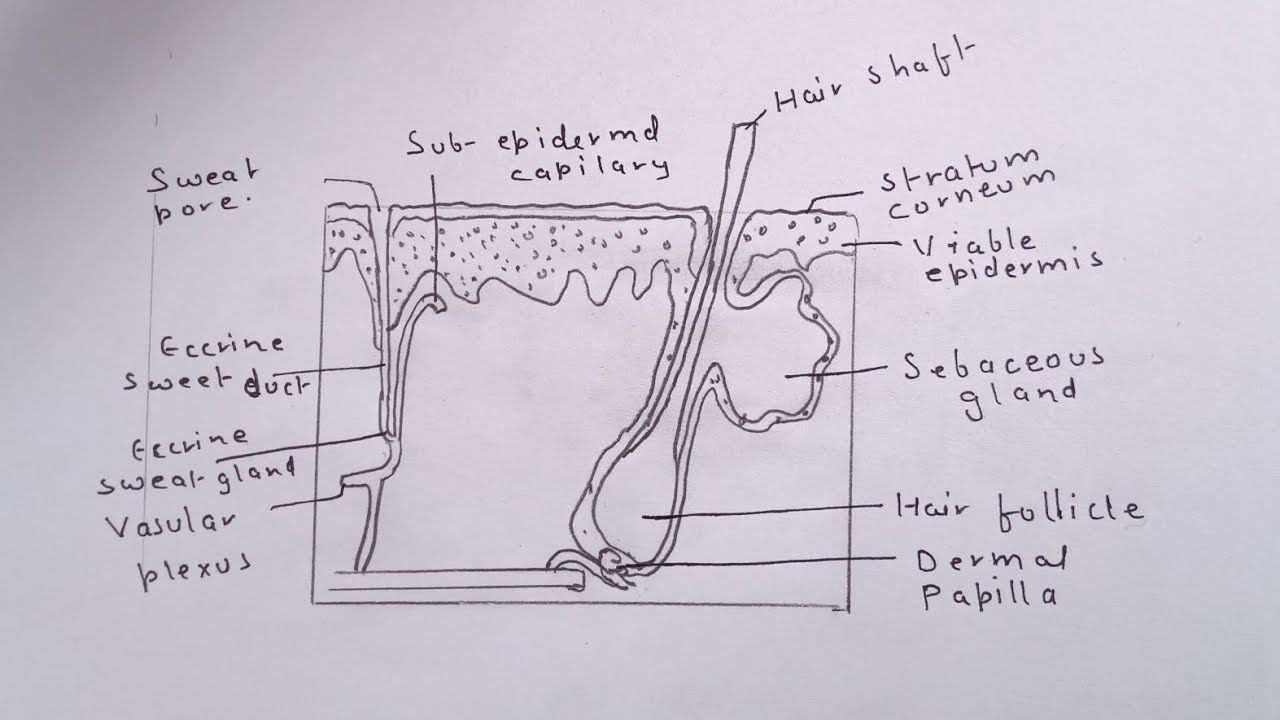
Human skin is similar to most of the other mammals' skin, and it is very similar to pig skin. Outer layer of the skin. Learn about the skin's function and conditions that may affect the skin. This skin diagram lists all the important parts of human skin, including the dermis, epidermis, hypodermis, sweat pore, hair shaft, pigment layer, nerve. Webmd's skin anatomy page provides a detailed image of the skin and its parts as well as a medical definition.
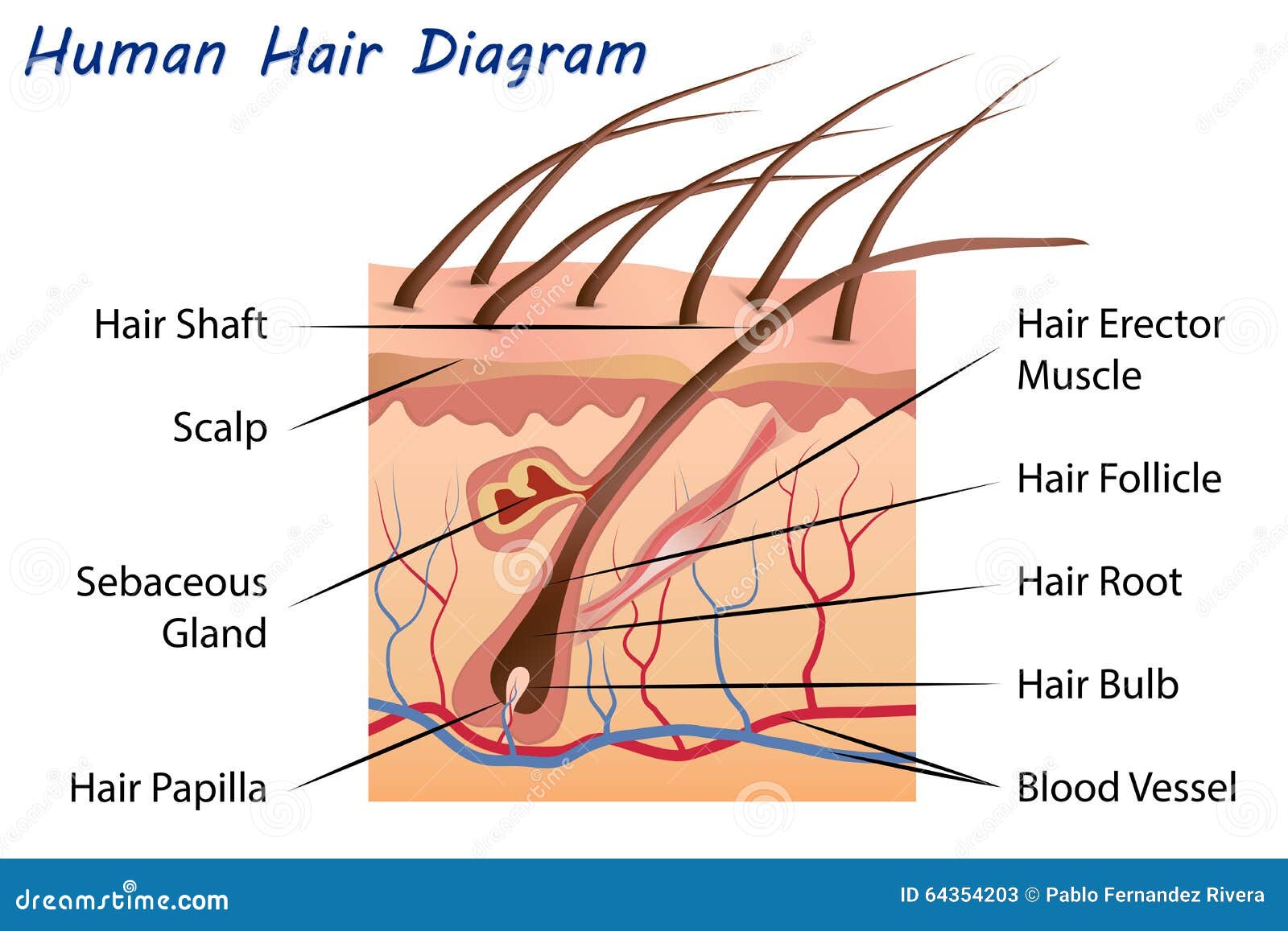
Human Hair Diagram Stock Illustrations 1 203 Human Hair Diagram Stock Illustrations Vectors Clipart Dreamstime from thumbs.dreamstime.com The thickness of skin varies from 0.5mm thick on the eyelids to 4.0mm thick on the heels of your feet. Skin is made up of two layers that cover a third fatty layer. This article will describe the anatomy and histology of the skin. Select from premium skin cell diagram of the highest quality. This skin diagram lists all the important parts of human skin, including the dermis, epidermis, hypodermis, sweat pore, hair shaft, pigment layer, nerve. Description from picture of human body with organs labeled pictures wallpaper : The outer layer is called the epidermis; Is a health blogger focusing on health, beauty, lifestyle and fitness topics.
Humans are miraculous beings, capable of doing anything they set their minds to.
ads/bitcoin2.txt
A keratinocyte is a cell that manufactures and stores the protein keratin. This skin diagram lists all the important parts of human skin, including the dermis, epidermis, hypodermis, sweat pore, hair shaft, pigment layer, nerve. He has been with healthiack.com since 2012 and has written and reviewed well over 500 coherent articles. In addition to human anatomy and physiology, this collection of images illustrates many of the major diseases and conditions of the body. This hd wallpaper picture of human body with organs labeled has viewed by 1017 users. Sensory receptors in the human skin. Literally covering you from head to toe. An average square inch of skin contains 650 sweat glands, 20 blood vessels, and more than 1,000 nerve endings. Skin has two main layers, both of which serve a purpose. The skin becomes dark color. Also explore over 33 similar quizzes in this category. A filament that grows from the skin. Middle layer of the skin.
Human skin is only about 0.07 inches (2 mm) thick. Posted in bones, diagrams | tagged body skeleton, human skeletal anatomy, human skeleton, human skeleton anatomy, skeletal, skeletal anatomy, skeletal images, skeletal system, skeleton picture of body organs location 2. Anatomy human body organs female 14 photos of the anatomy human body organs female anatomy human body appendix, anatomy human body liver, anatomy human body spleen, anatomy human body systems, anatomy of human body organs male, anatomy of the human body internal organs, diagram of human body organs in. Detailed texture of human skin. The cells in all of the layers except the stratum basale are called keratinocytes.
Structure And Function Of Skin Biology For Majors Ii from s3-us-west-2.amazonaws.com This skin diagram lists all the important parts of human skin, including the dermis, epidermis, hypodermis, sweat pore, hair shaft, pigment layer, nerve. Find the perfect skin cell diagram stock photos and editorial news pictures from getty images. It has an area of 2 square metres (22 square feet) in adults, and weighs about 5 kilograms. The epidermis, an outermost layer that contains the primary protective structure, the stratum corneum; The male was sectioned at one millimeter intervals Find free pictures, photos, diagrams, images and information related to the human body right here at science kids. The outer layer is called the epidermis; Select from premium skin cell diagram of the highest quality.
Also explore over 33 similar quizzes in this category.
ads/bitcoin2.txt
The epidermis, an outermost layer that contains the primary protective structure, the stratum corneum; Picture of human body with organs labeled, download this wallpaper for free in hd resolution.picture of human body with organs labeled was posted in june 11, 2017 at 5:58 am. The outer layer is called the epidermis; Oral health explore images of dental and oral health diseases as well as cosmetic dentistry before and after pictures. Skin also helps maintain a constant body temperature. Labelled pictures of human skin : Select from premium skin cell diagram of the highest quality. A filament that grows from the skin. Desquamation (sloughing of cells) from the epidermis, thick skin, human, 100x at 35mm. Find free pictures, photos, diagrams, images and information related to the human body right here at science kids. This diagram depicts labeled human skeleton diagram with parts and labels. Anatomy human body organs female 14 photos of the anatomy human body organs female anatomy human body appendix, anatomy human body liver, anatomy human body spleen, anatomy human body systems, anatomy of human body organs male, anatomy of the human body internal organs, diagram of human body organs in. When ultraviolet light waves touch melanocytes, they begin to increase the production of melanin.
ads/bitcoin3.txt
ads/bitcoin4.txt
ads/bitcoin5.txt
0 Response to "Labelled Pictures Of Human Skin : The Skin Human Anatomy Picture Definition Function And Skin Conditions"


0 Response to "Labelled Pictures Of Human Skin : The Skin Human Anatomy Picture Definition Function And Skin Conditions"
Post a Comment